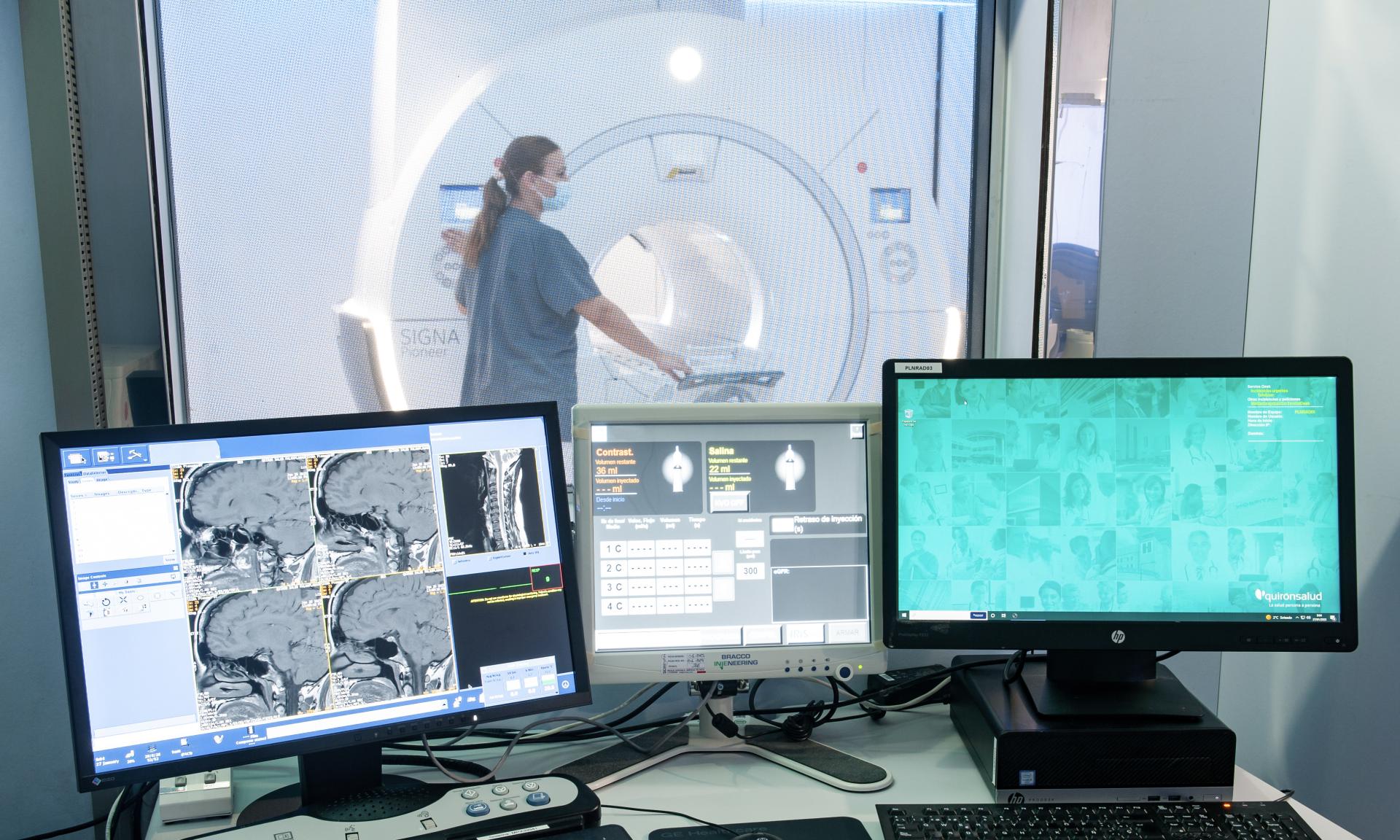
Artificial Intelligence, AI, is revolutionizing the diagnosis of pathologies and Hospital Quirónsalud Barcelona is a clear example of this.
Thanks to the addition of the Signa Voyager Air IQ Edition 1.5T MRI, the hospital has made a quantum leap in the quality of its diagnostics and patient care.
This new equipment has an image generation system based on a Deep Learning algorithm.
As a result, it is possible to obtain a sharper image and greater diagnostic accuracy, even in small structures such as those found intracranially.
For her part, Dr. Nadine Romero, head of the Diagnostic Imaging Unit at Hospital Quirónsalud Barcelona, emphasizes that “the result is more precise MRI images, achieving a highly reliable diagnostic result.
Quirónsalud Barcelona and the use of AI
In addition to artificial intelligence technology, the equipment also features specific sequences and software not found in other MRIs.
The ZTE sequences allow the analysis of bone structures and the specific software allows the assessment of myocardial strain or deformity, all without exposing the patient to ionizing radiation.
The application of artificial intelligence in medicine is becoming increasingly common and its impact is undeniable.
Algorithms that analyze patient data make it possible to provide more accurate diagnoses and personalized treatments.
In addition, AI can also detect incurable diseases from hidden data that could hide a condition.
In the case of Hospital Quirónsalud Barcelona, the incorporation of the Signa Voyager Air IQ Edition 1.5T MRI with Artificial Intelligence demonstrates the center’s commitment to offering the best medical care to its patients.
High technology in diagnostic imaging is a vital tool for the detection and treatment of pathologies. Thanks to AI, healthcare professionals can solve complex problems more effectively.
New diagnostic tools for more accurate treatments
Technology is advancing by leaps and bounds in medicine and with it, the ability to obtain accurate images of the inside of the human body.
These images are essential for disease diagnosis and medical decision making.
Magnetic resonance imaging (MRI), for example, is one of the most commonly used techniques to obtain these images.
New advances are being developed all the time to improve their application in healthcare.
Imaging complements MRI soft tissue examinations and offers a more accurate tool for the diagnosis of bone disease.
For cardiovascular studies, Siga Voyager 1.5T MRI provides reliable images of cardiac anatomy and function.

